


Genus Didemnum
This genus contains several North American native species which have been overshadowed by the spread of the invasive species Didemnum vexillum. The light micrographic specimens of Didemnum on this page were collected and processed in California during the early 1970s and labeled as Didemnum carnulentum. Digital color images were recorded beginning in Spring of 2011. Over the last 10 years, Didemnum vexillum has been extensively studied on a global basis resulting in scores of publications. The images I'll be including will hopefully be helpful to field biologists studying the spread of Didemnum vexillum as well as fill an untapped photographic and microscopic niche.
DIDEMNUM
Photomicroscopy and Anatomy Didemnum sp (carnulentum?)
Didemnum vexillum - cream-colored agressive invasive species
Botrylloides violaceus - maroon-colored nuisance species
Didemnum vexillum
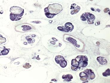
Cross section through the colony parallel to the surface - different zooids are sectioned through different planes. 5X obj.
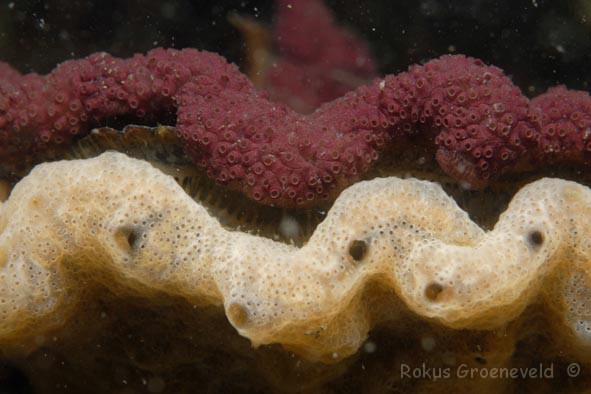
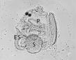
The Didemnum colony has a smooth, cream-colored surface marked by numerous tiny branchial openings and fewer larger cloacal apertures (stars). The cloacal chambers serve as an excurrent channel for the atrial opening of individual zooids as well as a brooding chamber for embryos. The surface of the tunic contains microscopic spiny calcareous spicules. Diplosoma has a very similar structure but does not posess spicules. Photo by Rokus Groeneveld www.diverosa.com
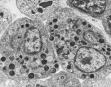
Cross section through the branchial basket showing endostyle and branchial basket.. 20X obj.
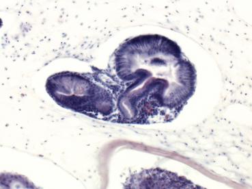
Cross section through the stomach (right) and intestine (left). 20X obj.
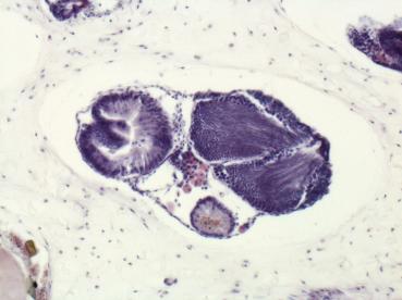
Cross section through the stomach (left) , intestine (small tube), and larger testis (right). 20X obj.
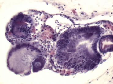
Cross section through two oocytes at different stages of developemnt (lower left) at the level of the stomach (right) and intestine (above larger oocyte). 40X obj.
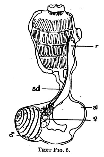
Relationship of oocytes and testis to stomach and intestine in Didemnum candidum. r, rectum; sd, sperm duct; st, stomach. (from Brewin, B.I., Ascidians in the Vicinity of the Portobello Marine Biological Station, Otago Harbour. Trans. Roy. Soc. New Zealand 76, 87-131, 1946.
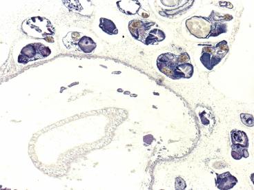
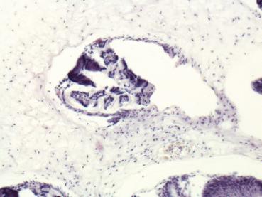
Cross section through the colony parallel to the surface showing the large cloacal chamber next to individual zooids. 5X obj.
Photographs and Stereozoom Microscopic images of Didemnum vexillum from Salem Sound, 2011 Didemnum vexillum
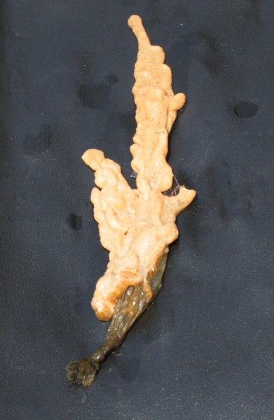
When present, Didemnum rapidly grows on flat surfaces of floating and subtidal structures and continues to spread over other species that provide a substrate. This includes mussels, sea grass, algae, and solitary ascidians. Many of the Styela clava seen during August were covered with Didemnum, and the only part of Styela that showed were the openings of the branchial and atrial siphons. On this specimen, Didemnum had completely grown over the top of Styela and was extending about 8 inches from the side of the dock. All that remained of the Styela was the lower portion of the tunic and the attachement site.
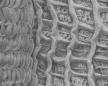
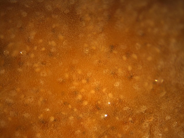
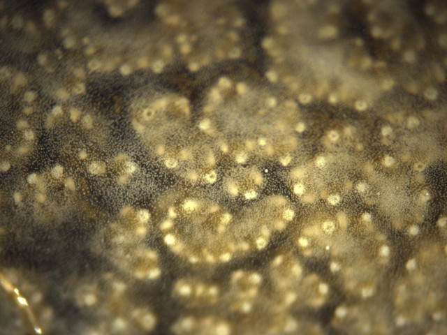
Stereozoom micrographs of the surface of live Didemnum vexillum showing groups of zooids, branchial siphons, and spicules. The top image was taken of a specimen a few mm thick colored similarly to the specimen at the left. The image below is of a single layer of a colony growing over a mussel shell, which provided a dark, contrasting background to the Didemnum. Both images taken using 3X zoom setting.