


STYELA
Styela clava
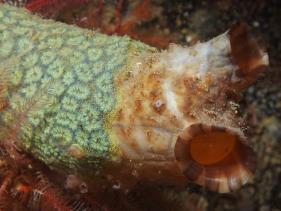
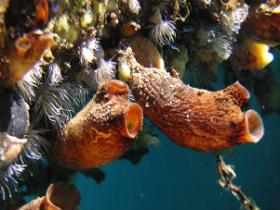
Photographic Credits: clockwise from upper left, 1, Charlie Waters. 2, Janna Nichols. 3, Danuel Guip's Photostream, 4. Doug Mason's Photostream, Botryllus schlosseri on Styela clava.
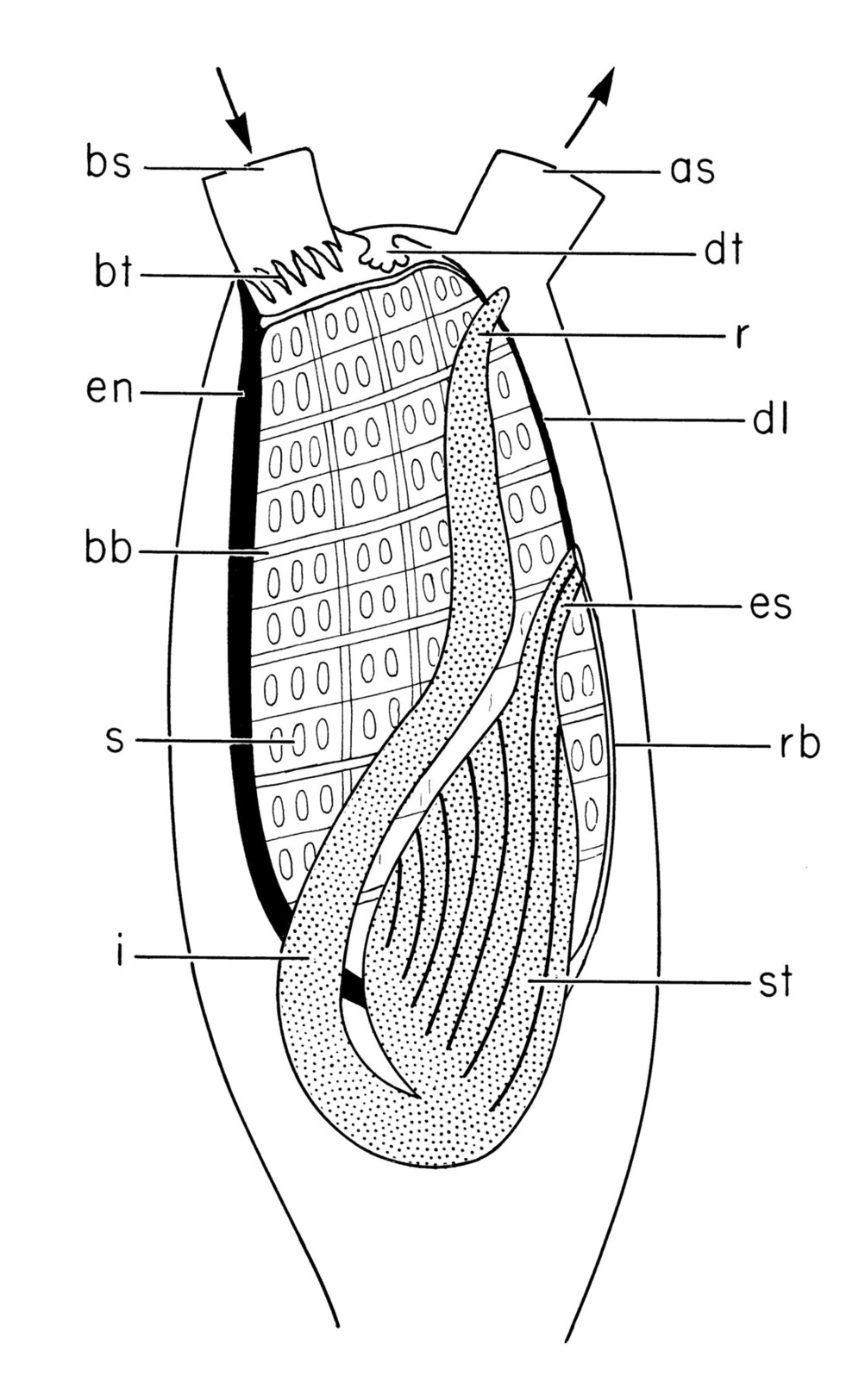
GI Tract of Styela clava. Abbreviations: bs, branchial siphon, bt, branchial tentacles; en, endostyle; bb, branchial bar; s, stigmata; dt, dorsal tubercle; dl, dorsal lamina; rb, retropharyngeal band; es, esophagus; st, stomach; i, intestine; r, rectum; as, atrial siphon. Arrows indicate direction of water flow. Original artwork, Ermak, 1975.
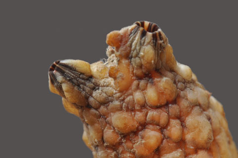
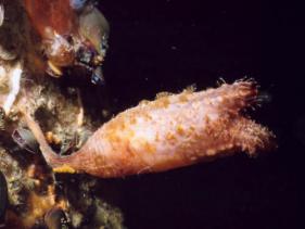
Styela clava with siphons almost closed. Closed siphones form cones. Open siphons are flared outwards at the ends, like photo #3 above. Web photo from nathistoc.bio.uci.edu Phototaken at HMB docks
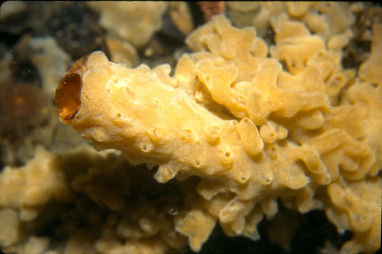
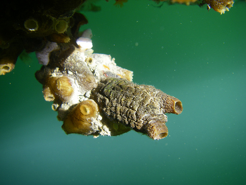
Common view of Styela clava in late summer 2011 completely covered with Didemnum vexillus except for the ends of the atrial siphons. On most of the specimens I checked out, the Didemnum was not attached to the siphons and the siphons could expand and contract freely. Woods Hole web photo.
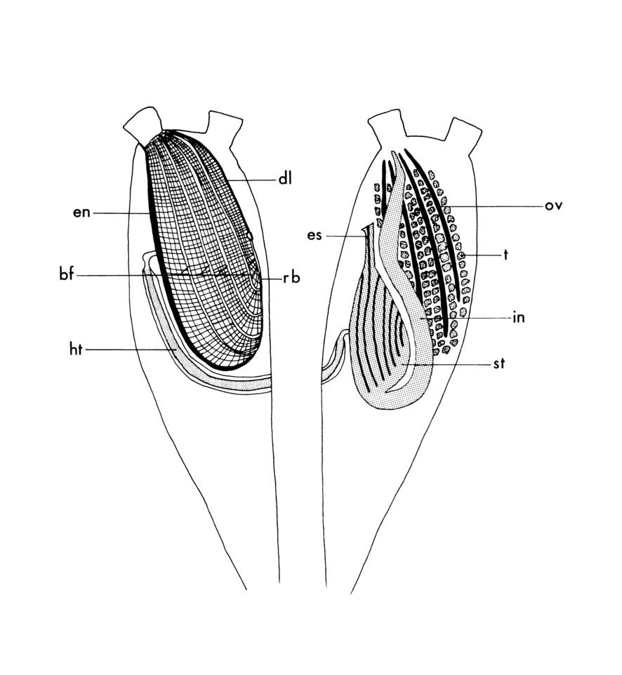
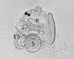
Anatomy of Styela clava showing reproductive organs (right side only), digestive tract, and heart. en, endostyle; bf, branchial folds; ht, heart; dl, dorsal lamina; rb, retropharyngeal band; es, esophagus; ov, ovary; t, testes; in, intestine; st, stomach. Ermak, T.H. Ph.D. Thesis. University of California, San Diego; Scripps Institution of Oceanography Library. Fig. 1, p. 19, 1975.